Tuesday, 27 September 2016

Common blood collection tubes, their additives and laboratory uses

The evacuated tube system for blood collection in use for various laboratory tests consists of tubes of various sizes, with color coded tops indicating tube contents. Most blood collection tubes contain an additive that either accelerates clotting of the blood (clot activator) or prevents the blood from clotting (anticoagulant).
The list below lists the most commonly used blood collection tubes, their additives and uses in laboratory:
1. Red
The red bottle is less common – it is used for biochemistry tests requiring serum which might be adversely affected by the separator gel used in the yellow bottle.
Additive: None or contains silica particles which act as clot activators.
What additive does: Clot activator promotes blood clotting with glass or silica particles.
Laboratory Uses: Serum testing (glucose, cholesterol, triglycerides, HDL, potassium, amylase, alkaline phosphatase, BUN, CK, liver enzymes), blood bank, serology (RH Typing, Antibody screening, Red Cell Phototyping, DAT, RPR, monospot, rheumatoid factor, ANA)
2. Yellow
Additive: anticoagulant SPS (Sodium Polyanetholsulfonate) & ACD (acid citrate dextrose)
What additive does: Prevents the blood from clotting and stabilizes bacterial growth.
Laboratory Uses: Blood and bodily fluid cultures (HLA, DNA, Paternity)
Tubes with SPS – For Blood and bodily fluid cultures (HLA, DNA, Paternity). The SPS aids in the recovery of microorganisms by slowing down/ stopping the actions of complement, phagocytes, and certain antibiotics.
Tubes with ACD are for cellular studies, HLA typing, paternity testing.
3. Light Blue
The blue bottle is used for haematology tests involving the clotting system, which require inactivated whole blood for analysis.
Additive: Sodium Citrate
What additive does: Binds and remove calcium to prevent blood from clotting
Laboratory uses: Coagulation (clotting process-P.T)
PT (Prothrombin Time – evaluates the extrinsic system of the coagulation cascade & monitors coumadin therapy)
APTT/ PTT (Activated Partial Thromboplastin Time – evaluates the intrinsic system of the coagulation cascade & monitors heparin therapy)
FDP (Fibrinogen Degradation Products)
TT (Thrombin Time)
Factor assays
4. Green
This less commonly used bottle is for biochemistry tests which require heparinised plasma or whole blood for analysis.
Additive: Heparin (Sodium/Lithium/Ammonium)
What additive does: Inhibits thrombin formation to prevent clotting
Laboratory uses: Chemistry Testing (Plasma determinations in chemistry) : ammonia, carboxyhemoglobin & STAT electrolytes, chromosome screening, insulin, renin and aldosterone
5. Lavender
These bottles are generally used for haematology tests where whole blood is required for analysis.
Additive: EDTA (Ethylenediaminetetraacetic Acid)
What additive does: Removes calcium preventing clotting of blood
Laboratory uses: Hematology testing (ESR, CBC w/diff., HgBA1c)
blood film for abnormal cells or malaria parasites, reticulocytes, red cell folate, Monospot test for EBV, parathyroid hormone (PTH)
6. Grey
Additive: Potassium oxalate and Sodium fluoride
What additive does: Sodium fluoride acts as an antiglycolytic agent to ensure that no further glucose breakdown occurs within the sample after it is taken. Potassium oxalate removes calcium and acts as an anticoagulant.
Laboratory uses: Chemistry testing, especially glucose(sugar) and lactate, Glucose tolerance test (GTT)
7. Royal Blue
Additive: Sodium Heparin also Sodium EDTA
What additive does: Inhibits Thrombin formation to prevent
Laboratory uses: Chemistry trace elements (such as Zinc, Copper, Lead and Mercury), toxicology, and nutritional chemistry testing
8. Black
Additive: Sodium citrate
what additive does: Forms calcium salts to remove calcium
Laboratory uses: paediatric ESR

Monday, 26 September 2016

Hormonal Profile

If you are unable to become pregnant after one year of unprotected intercourse, or six months of unprotected intercourse if you are a woman over 35, you and your partner should have a full physical and medical evaluation.

Several blood tests are available to determine if you, your partner, or both of you have a problem that is causing infertility.

FSH

Follicle-stimulating hormone (FSH) helps control a woman’s menstrual cycle and the production of eggs. In men, it regulates the production and transportation of sperm.

For women, a FSH test is done on the third day of the menstrual cycle and is used to evaluate egg supply. For men, the test is used to determine sperm count.

Estradiol

Estradiol is an important form of estrogen. Anestradiol test is used to measure a woman’s ovarian function and to evaluate the quality of the eggs. Like FSH, it is done on the third day of a woman’s menstrual cycle.

Luteinizing Hormone Level

In women, luteinizing hormone (LH) is linked to ovarian hormone production and egg maturation. In men, it stimulates the hormone testosterone which affects sperm production.

An LH test is used to measure a woman’s ovarian reserve (egg supply) and a man’s sperm count. It is done during a woman's menstrual cycle to see if she is ovulating

Serum Progesterone

Progesterone is a female hormone produced by the ovaries during ovulation. It causes the endometrial lining of the uterus to get thicker, making it receptive for a fertilized egg.

A serum progesterone test is used to determine if ovulation is occurring. Sinceprogesterone levels increase towards the end of a woman’s cycle, the test is done during the luteal phase of the menstrual cycle (just before her period starts).

Prolactin

The hormone prolactin is made by the pituitary gland and causes milk production.

In women, a prolactin test is done to find out why they are not menstruating, or why they are having infertility problems or abnormal nipple discharge. The test is done in men when there is a lack of sexual desire, difficulty getting an erection, or if there might be a problem with the pituitary gland.

Androgen

Testosterone is probably the most well-known androgen and it affects the sexual functioning of both men and women.

In men, an androgen test is used to find the cause of a low sex drive, the inability to get an erection, or infertility. In women, it is used to determine the cause of irregular periods or a low sex drive.

Cross-matching

Cross Matching is a procedure performed prior to a blood transfusion to determine whether donor blood is compatible (or incompatible) with recipient blood. Compatibility is determined through matching of different blood group systems, the most important of which are the ABO and Rh system, and/or by directly testing for the presence of antibodies against a sample of donor tissues or blood.

Purpose of Cross Matching

The crossmatch is routinely used as the final step of pretransfusion compatibility testing. The purposes of compatibility testing are to detect: irregular antibodies; errors in ABO grouping, and clerical errors in patient identification and result recording. The crossmatch will detect the following:
1. Most recipient antibodies directed against antigens on the donor red blood cells.
2. Major errors in ABO grouping, labeling, and identification of donors and recipients.

Principle

Cross-matching will detect incompatibilities between the donor and recipient that will not be evident on blood typing. There are two types of cross-matches: Major cross-match and Minor cross-match.

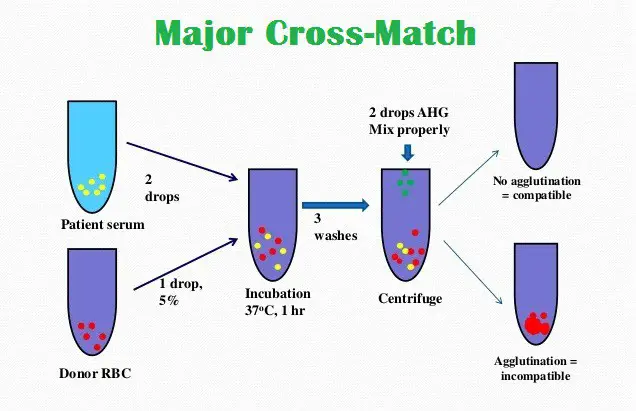
The major crossmatch involves testing the patient’s serum with donor cells to determine whether the patient has an antibody which may cause a hemolytic transfusion reaction or decreased cell survival of donor cells. This is the most important cross-match.

The minor crossmatch involves testing the patients cells with donor plasma to determine whether there is an antibody in the donor’s plasma directed against an antigen on the patient’s cells.

Procedure

Prepare donor and recipient blood samples:
For Major crossmatch : Donor’s red cell and recipient serum or plasma
For Minor crossmatch : Recipient red cells and donor’s serum or plasmaPrepare 3 – 5% cell suspensions of red cells.Major Crossmatch:
Label a test tube. Add two drops of the patient serum and one drop of the appropriate donor cell suspension.Minor Crossmatch:
Label a test tube. Add two drops of the appropriate donor serum and one drop of the patient cell suspension.Mix the tubes and incubate at 37°C for about 45 minutes.Add two drops of AHG (Antihuman globulin) and mix well.Centrifuge for 1 minute at 1500 rpmRead macroscopically and microscopically and record the results

Interpretation

The mixture of erythrocytes and serum are observed for hemolysis or microscopically for agglutination. Any evidence of hemolysis/agglutination indicates an incompatible cross-match. Negative results are taken to indicate compatibility.

Blood group

Blood Types

Human blood is grouped into four types: A, B, AB, and O. Each letter refers to a kind of antigen, or protein, on the surface of red blood cells. For example, the surface of red blood cells in Type A blood has antigens known as A-antigens.

The Rh Factor

Each blood type is also grouped by its Rhesus factor, or Rh factor. Blood is either Rh positive (Rh+) or Rh negative (Rh-). About 85% of Americans have Rh+ blood.

Rhesus refers to another type of antigen, or protein, on the surface of red blood cells. The name Rhesus comes from Rhesus monkeys, in which the protein was discovered.

Blood Transfusions

Blood types become very important when a blood transfusion is necessary. In a blood transfusion, a patient must receive a blood type that is compatible with his or her own blood type—that is, the donated blood must be accepted by the patient's own blood. If the blood types are not compatible, red blood cells will clump together, making clots that can block blood vessels and cause death.

Type O– blood is considered the “universal donor” because it can be donated to people of any blood type. Type AB+ blood is considered the “universal recipient” because people with this type can receive any blood type.

Sunday, 25 September 2016

Blood Transfusion

Definition

Transfusion is the process of transferring whole blood or blood components from a donor to a recipient.

Purpose

Transfusions are given to restore lost blood, to improve clotting time, and to improve the ability of the blood to deliver oxygen to the body's tissues. About 32,000 pints of donated blood are transfused each day in the Nigeria.

The three main reasons why a child may need a blood transfusion are:

Loss of blood during surgery or from an injury or an illness.An inability to make enough blood.Some illnesses and treatments can harm the bone marrow's ability to make blood (e.g., chemotherapydecreases production of new blood cells).To prevent complications from an existing blood or bleeding disorder, such as sickle cell disease, thalassemia, or anemia caused bykidney diseasehemophilia, or von Willebrand disease.

Where the Blood Comes From

Because there's no manmade substitute for blood, the blood supply used for transfusion must be donated. The three types of blood donation are:

Autologous (ah-TOL-uh-gus) blood donation. Sometimes, when people know in advance that they are going to need a transfusion (for a planned surgery, for example), they may donate their own blood beforehand. There's no age requirement, but in general, kids don't donate their blood for their own use until they're over age 12.
Directed donation. This is when a family member or friend with a compatible blood type donates blood specifically for use by a designated patient.
Volunteer donation. Since there's no medical evidence that blood from directed donors is any safer than blood from volunteer donors, most patients receive blood donated through blood drives, which are often run by independent collection agencies like the American Red Cross. The minimum age for donating blood is 16 or 17 years old, depending on where a person lives.

Some people worry about getting diseases from infected blood, but the United States has one of the safest blood supplies in the world. Many organizations, including community blood banks and the federal government, work hard to make sure that the blood supply is safe.

The risk of getting a disease like HIV or hepatitis through a transfusion is extremely low in the Nigeria today because of very stringent blood screening. Also, the needles and other equipment used are sterile, and are used only on one person and then thrown away in special containers.

Description

Blood is collected from the donor by inserting a large needle into a vein in the arm, usually one of the larger veins near the inside of the elbow. A tourniquet is placed on the upper arm to increase the pressure in the arm veins, which makes them swell and become more accessible. Once the nurse or technician has identified a suitable vein, she or he sterilizes the area where the needle will be inserted by scrubbing the skin with a soap solution or an antiseptic that contains iodine. Sometimes both solutions are used. The donor lies on a bed or cot during the procedure, which usually takes between 10 and 20 minutes. Generally, an 18-gauge needle is used. This size of needle fits easily into the veins and yet is large enough to allow blood to flow easily. Human blood will sometimes clot in a smaller needle and stop flowing. The donor's blood is collected in a sterile plastic bag that holds one pint (450 ml). The bags contain an anticoagulant to prevent clotting and preservatives to keep the blood cells alive. A sample of the donator's blood is collected at the time of donation and tested for infectious diseases. The blood is not used until the test results confirm that it is safe. Properly handled and refrigerated, whole blood can last for 42 days.

The recipient of a transfusion is prepared in much the same way as the blood donor. The site for the needle insertion is carefully washed with a soap-based solution followed by an antiseptic containing iodine. The skin is then dried and the transfusion needle inserted into the vein. During the early stages of a transfusion, the recipient is monitored closely to detect any adverse reactions. If no signs of adverse reaction are evident, the patient is monitored occasionally for the duration of the transfusion period. Upon completion of the transfusion, a compress is placed over the needle insertion site to prevent extensive bleeding.

Blood typing

All donated blood is typed, which means that it is analyzed to determine which of several major and minor blood types (also called blood groups) it belongs to. Blood types are genetically determined. The major types are classified by the ABO system. This system groups blood with reference to two substances in the red blood cells called antigen A and antigen B. The four ABO blood types are A, B, AB, and O. Type A blood has the A antigen, type B has the B antigen, type AB has both, and type O has neither. These four types of blood are further classified by the Rh factor. The Rh, or rhesus factor, is also an antigen in the red blood cells. A person who has the Rh factor is Rh positive; a person who does not have the factor is Rh negative. If a person has red blood cells with both the B and the Rh antigens, that person is said to have a B positive (B+) blood type. Blood types determine which kinds of donated blood a patient can receive. Generally, patients are limited to receiving only blood of the exact same ABO and Rh type as their own. For example, a person with B+ blood can receive blood or blood cells only from another person with B+ blood. An exception is blood type O. Individuals with type O blood are called universal donors, because people of all blood types can accept their blood.

Blood can also be typed with reference to several other minor antigens, such as Kell, Kidd, Duffy, and Lewis. These minor antigens can become important when a patient has received many transfusions. These patients tend to build up an immune response to the minor blood groups that do not match their own. They may have an adverse reaction upon receiving a transfusion with a mismatched minor blood group. A third group of antigens that may cause a reaction are residues from the donor's plasma attached to the RBCs. To eliminate this problem, the RBCs are rinsed to remove plasma residues. These rinsed cells are called washed RBCs.

Other transfusion procedures

Autologous transfusion is a procedure in which patients donate blood for their own use. Patients who are to undergo surgical procedures requiring a blood transfusion may choose to donate several units of blood ahead of time. The blood is stored at the hospital for the patient's exclusive use. Autologous donation assures that the blood type is an exact match. It also assures that no infection will be transmitted through the blood transfusion. Autologous donation accounts for 5% of blood use in the United States each year.

Directed donors are family or friends of the patient who needs a transfusion. Some people think that family and friends provide a safer source of blood than the general blood supply. Studies do not show that directed donor blood is any safer. Blood that is not used for the identified patient becomes part of the general blood supply.

Apheresis is a special procedure in which only certain specific components of a donor's blood are collected. The remaining blood fractions are returned to the donor. A special blood-processing instrument is used in apheresis. It fractionates the blood, saves the desired component, and pumps all the other components back into the donor. Because donors give only part of their blood, they can donate more frequently. For example, people can give almost ten times as many platelets by apheresis as they could give by donating whole blood. The donation process takes about one to two hours.

Preparation

The first step in blood donation is the taking of the donor's medical history. Blood donors are questioned about their general health, their lifestyle, and any medical conditions that might disqualify them. These conditions include hepatitis, AIDS, cancer, heart disease, asthma, malaria, bleeding disorders, and high blood pressure. Screening prevents people from donating who might transmit diseases or whose medical condition would place them at risk if they donated blood. Some geographical areas or communities have a high rate of hepatitis or AIDS. Blood collection in most of these areas has been discontinued indefinitely.

The blood pressure, temperature, and pulse of donors are taken to ensure that they are physically able to donate blood. One pint (450 mL) of blood is usually withdrawn, although it is possible to donate smaller amounts. The average adult male has 10–12 pints of blood in his body; the average adult female has 8–9 pints in hers. Within hours after donating, most people's bodies have replaced the fluid lost with the donated blood, which brings their blood volume back to normal. Replacement of the blood cells and platelets, however, can take several weeks. Pregnant women and people with low blood pressure or anemia should not donate blood or should limit the amount of blood they give. Generally, people are allowed to donate blood only once every two months. This restriction ensures the health of the donor and discourages people from selling their blood. The former practice of paying donors for blood has essentially stopped. Donors who sell blood tend to be at high risk for the transmission of bloodborne diseases.

Aftercare

Recipients of blood transfusion are monitored during and after the transfusion for signs of an adverse reaction. Blood donors are generally given fluids and light refreshments to prevent such possible side effects as dizziness and nausea. They are also asked to remain in the donation area for 15–20 minutes after giving blood to make sure that they are not likely to faint when they leave.

Risks

Risks for donors

For donors, the process of giving blood is very safe. Only sterile equipment is used and there is no chance of catching an infection from the equipment. There is a slight chance of infection at the puncture site if the skin is not properly washed before the collection needle is inserted. Some donors feel lightheaded when they sit up or stand for the first time after donating. Occasionally, a donor will faint. Donors are encouraged to drink plenty of liquids to replace the fluid lost with the donated blood. It is important to maintain the fluid volume of the blood so that the blood pressure will remain stable. Strenuous exerciseshould be avoided for the rest of the day. It is normal to feel some soreness or to find a small bluish bruise at the site of the needle insertion. Most donors have very slight symptoms or no symptoms at all after giving blood.

Risks for recipients

A number of precautions must be taken for transfusion recipients. Donated blood must be matched with the recipient's blood type, as incompatible blood types can cause a serious adverse reaction (transfusion reaction). Blood is introduced slowly by gravity flow directly into the veins (intravenous infusion) so that medical personnel can observe the patient for signs of adverse reactions. People who have received many transfusions may develop an immune response to some factors in foreign blood cells (see below). This immune reaction must be evaluated before the patient is given new blood.

Adverse reactions to mismatched blood (transfusion reaction) is a major risk of blood transfusion. Transfusion reaction occurs when antibodies in the recipient's blood react to foreign blood cells introduced by the transfusion. The antibodies bind to the foreign cells and destroy them. This destruction is called a hemolytic reaction. In addition, a transfusion reaction may also cause a hypersensitivity of the immune system that may in turn result in tissue damage within the patient's body. The patient may also have an allergic reaction to mismatched blood.

The first symptoms of transfusion reaction are a feeling of general discomfort and anxiety. Breathing difficulties, flushing, and a sense of pressure in the chest or back pain may also be present. Evidence of a hemolytic reaction can be seen in the urine, which will be colored from the hemoglobinleaking from the destroyed red blood cells. Severe hemolytic reactions are occasionally fatal. Reactions to mismatches of minor factors are milder. These symptoms include itchiness, dizziness, fever, headache, rash, and swelling. Sometimes the patient will experience breathing difficulties and muscle spasms. Most adverse reactions from mismatched blood are not life-threatening.

Infectious diseases can also be transmitted through donated blood and constitute another major risk of blood transfusion. The infectious diseases most often acquired from blood transfusion in the United States are hepatitis and HIV.

Patients who are given too much blood can develop high blood pressure, a concern for people who have heart disease. Very rarely, an air embolism is created when air is introduced into a patient's veins through the tubing used for intravenous infusion. The danger of embolism is greatest when infusion is begun or ended. Care must be taken to ensure that all air is bled out of the tubing before infusion begins, and that the infusion is stopped before air can enter the patient's blood system.

Normal results

Most individuals will feel only a slight sting from the needle used during the blood donation process, and will not experience any side effects after the procedure is over. Plasma is regenerated by the body within 24 hours, and red blood cells within a few weeks. Patients who receive a blood transfusion will usually experience mild or no side effects.

Morbidity and mortality rates

The risk of acquiring an infectious disease from a blood transfusion is very low. The risk of HIV transmission is one in 450,000 to 660,000 units of blood; hepatitis B virus (HBV), one in 137,000 units; and hepatitis C virus (HCV), one in 1,000,000 units. Bacterial contamination (a cause of infection) is identified in one in 4,200 transfusions. Approximately one in 25,000 individuals who receive a blood transfusion will develop a hemolytic reaction; the risk of a fatal hemolytic reaction is one in 160,000.

Alternatives

There are several alternatives to blood transfusion as of 2003. These include:

Volume expanders. Certain fluids (saline, Ringer's lactate solution, dextran, etc.) may be used to increase the patient's blood volume without adding additional blood cells.Blood substitutes. Much research is currently being done into compounds that can replace some or all of the functions of blood components. One such compound, called HBOC-201 or Hemopure, is hemoglobin derived from bovine (cow) blood. Hemopure shows promise as a substitute for red blood cell transfusion.Bloodless surgery. It may be possible to avoid excessive blood loss through careful planning prior to surgery. Specialized instruments can minimize the amount of blood lost during a procedure. It is also possible to collect some of the blood lost during surgery and reinfuse it into the patient at the end of the operation.

Packed Cell Volume

Definition

The hematocrit is a test that measures the percentage of blood that is comprised of red blood cells.

Purpose

The hematocrit is used to screen for anemia, or is measured on a person to determine the extent of anemia. Ananemic person has fewer or smaller than normal red blood cells. A low hematocrit, combined with other abnormal blood tests, confirms the diagnosis. The hematocrit is decreased in a variety of common conditions including chronic and recent acute blood loss, some cancers, kidney and liver diseases, malnutrition, vitamin B 12and folic acid deficiencies, iron deficiency, pregnancy, systemic lupus erythematosus, rheumatoid arthritis and peptic ulcer disease. An elevated hematocrit is most often associated with severe burns, diarrhea, shock, Ad dison's disease, and dehydration, which is a decreased amount of water in the tissues. These conditions reduce the volume of plasma water causing a relative increase in RBCs, which concentrates the RBCs, called hemoconcentration. An elevated hematocrit may also be caused by an absolute increase in blood cells, called polycythemia. This may be secondary to a decreased amount of oxygen, called hypoxia, or the result of a proliferation of blood forming cells in the bone marrow (polycythemia vera).


Critically high or low levels should be immediately called to the attention of the patient's nurse or doctor.Transfusion decisions are based on the results of laboratory tests, including the hematocrit. Generally, transfusion is not considered necessary if the hematocrit is above 21%. The hematocrit is also used as a guide to how many transfusions are needed. Each unit of packed red blood cells administered to an adult is expected to increase the hematocrit by approximately 3% to 4%.


Precautions

Fluid volume in the blood affects hematocrit values. Accordingly, the blood sample should not be taken from an arm receiving IV fluid or during hemodialysis. It should be noted that pregnant women have extra fluid, which dilutes the blood, decreasing the hematocrit. Dehydration concentrates the blood, which increases the hematocrit.

In addition, certain drugs such as penicillin and chloramphenicol may decrease the hematocrit, while glucose levels above 400 mg/dL are known to elevate results. Blood for hematocrit may be collected either by finger puncture, or sticking a needle into a vein, called venipuncture. When performing a finger puncture, the first drop of blood should be wiped away because it dilutes the sample with tissue fluid. A nurse or phlebotomist usually collects the sample following cleaning and disinfecting the skin at the site of the needle stick.


Description

Blood is made up of red blood cells, white blood cells (WBCs), platelets, and plasma. A decrease in the number or size of red cells also decreases the amount of space they occupy, resulting in a lower hematocrit. Conversely, an increase in the number or size of red cells increases the amount of space they occupy, resulting in a higher hematocrit. Thalassemia minor is an exception in that it usually causes an increase in the number of red blood cells, but because they are small, it results in a decreased hematocrit.

The hematocrit may be measured manually by centrifugation. A thin capillary tube called a microhematocrit tube is filled with blood and sealed at the bottom. The tube is centrifuged at 10,000 RPM (revolutions per minute) for five minutes. The RBCs have the greatest weight and are forced to the bottom of the tube. The WBCs and platelets form a thin layer, called the buffy coat, between the RBCs and the plasma, and the liquid plasma rises to the top. The height of the red cell column is measured as a percent of the total blood column. The higher the column of red cells, the higher the hematocrit. Most commonly, the hematocrit is measured indirectly by an automated blood cell counter. It is important to recognize that different results may be obtained when different measurement principles are used. For example, the microhematocrit tube method will give slightly higher results than the electronic methods when RBCs of abnormal shape are present because more plasma is trapped between the cells.


Aftercare

Discomfort or bruising may occur at the puncture site. Pressure to the puncture site until the bleeding stops reduces bruising; warm packs relieve discomfort. Some people feel dizzy or faint after blood has been drawn, and lying down and relaxing for awhile is helpful for these people.


Risks

Other than potential bruising at the puncture site, and/or dizziness, there are no complications associated with this test.


Normal results

Normal values vary with age and sex. Some representative ranges are:

  • at birth: 42-60%
  • six to 12 months: 33-40%
  • adult males: 42-52%
  • adult females: 35-47%


Phlebotomy

Definition

Phlebotomy is the act of drawing or removing blood from the circulatory system through a cut (incision) or puncture in order to obtain a sample for analysis and diagnosis. Phlebotomy is also done as part of the patient's treatment for certain blood disorders.

Purpose

Phlebotomy that is part of treatment (therapeutic phlebotomy) is performed to treat polycythemia vera, a condition that causes an elevated red blood cell volume ( hematocrit ). Phlebotomy is also prescribed for patients with disorders that increase the amount of iron in their blood to dangerous levels, such as hemochromatosis, hepatitis B, and hepatitis C. Patients with pulmonary edema may undergo phlebotomy procedures to decrease their total blood volume.

Phlebotomy is also used to remove blood from the body during blood donation and for analysis of the substances contained within it.

Description

Phlebotomy is performed by a nurse or a technician known as a phlebotomist. Blood is usually taken from a vein on the back of the hand or just below the elbow. Some blood tests, however, may require blood from an artery. The skin over the area is wiped with an antiseptic, and an elastic band is tied around the arm. The band acts as a tourniquet, retaining blood within the arm and making the veins more visible. The phlebotomy technician feels the veins in order to select an appropriate one. When a vein is selected, the technician inserts a needle into the vein and releases the elastic band. The appropriate amount of blood is drawn and the needle is withdrawn from the vein. The patient's pulse and blood pressure may be monitored during the procedure.

For some tests requiring very small amounts of blood for analysis, the technician uses a finger stick. A lance, or small needle, makes a small cut in the surface of the fingertip, and a small amount of blood is collected in a narrow glass tube. The fingertip may be squeezed to get additional blood to surface.

The amount of blood drawn depends on the purpose of the phlebotomy. Blood donors usually contribute a unit of blood (500 mL) in a session. The volume of blood needed for laboratory analysis varies widely with the type of test being conducted. Typically one or several small (5–10 mL) tubes are drawn. Therapeutic phlebotomy removes a larger amount of blood than donation and blood analysis require. Phlebotomy for treatment of hemochromatosis typically involves removing a unit of blood—250 mg of iron—once a week. Phlebotomy sessions are required until iron levels return to a consistently normal level, which may take several months to several years. Phlebotomy for polycythemia vera removes enough blood to keep the patient's hematocrit (proportion of red blood cells) below 45%. The frequency and duration of sessions depends on the patient's individual needs.

Diagnosis/Preparation

Patients having their blood drawn for analysis may be asked to discontinue medications or to avoid food (to fast) for a period of time before the blood test. Patients donating blood will be asked for a brief medical history, have their blood pressure taken, and have their hematocrit checked with a finger stick test prior to donation.

Aftercare

After blood is drawn and the needle is removed, pressure is placed on the puncture site with a cotton ball to stop bleeding, and a bandage is applied. It is not uncommon for a patient to feel dizzy or nauseated during or after phlebotomy. The patient may be encouraged to rest for a short period once the procedure is completed. Patients are also instructed to drink plenty of fluids and eat regularly over the next 24 hours to replace lost blood volume. Patients who experience swelling of the puncture site or continued bleeding after phlebotomy should seek immediate medical treatment.

Risks

Most patients will have a small bruise or mild soreness at the puncture site for several days. Therapeutic phlebotomy may cause thrombocytosis and chronic iron deficiency (anemia) in some patients. As with any invasive procedure, infection is also a risk. This risk is minimized by the use of prepackaged sterilized equipment and careful attention to proper technique. There is no risk of HIV infection from phlebotomy, since all needles are disposed of after a single use. Arterial blood collection carries a higher risk than venous collection, and is performed by a physician or other specially trained professional. Patients who are anemic or have a history of cardiovascular disease may not be good candidates for phlebotomy.

Normal results

Normal results include obtaining the needed amount of blood with the minimum of discomfort to the patient.

Morbidity and mortality rates

Properly performed, phlebotomy does not carry the risk of mortality. It may cause temporary pain and bleeding, but these are usually easily managed.

Alternatives

Phlebotomy is a necessary medical procedure, and is required for a wide variety of other procedures.